Looking for the information on Gel Electrophoresis? Here, in this blog, you will get answers to all questions related to Gel Electrophoresis!
Definition of Gel Electrophoresis
A technique that is used to separate mixtures of nucleic acids such as DNA, RNA and even proteins according to their molecular size is Gel electrophoresis! Let’s take a dive into its introduction.
This separation technique is used in basic biotechnology lab as well as in the domain of clinical chemistry so as to separate proteins according to their charge or size and in biochemistry and molecular biology, a mixed population of DNA and RNA fragments could be separated by length.
It is also used to estimate the size of DNA and RNA fragments by comparing the gel picture of separated DNA fragments with the preloaded DNA ladder. Commercial DNA ladders usually come in different size ranges, so we need to pick the one with good “coverage” of the size range of our expected fragments.
Size Independent Gel Electrophoresis
In certain cases size independent gel electrophoresis using IEF (Isoelectric Focusing) agarose is run. The whole separation process takes place under the influence of magnetic field and its effects.
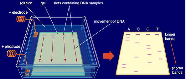
In this separation technique, the molecules which are to be separated are pushed by an electrical field through a gel. The gel is porous in nature and contains small pores through which the molecules travel. The travelling speed of the molecules is inversely proportional to the length of the molecules.
That means large DNA fragments tend to travel slowly as compared to the small DNA segments through the gel. The matrix of the gel like agar or polyacrylamide is the medium through which a charge is applied as along with the gel matrix the gel is immersed in the buffer which becomes the conducting medium due to the presence of ions in it.
The biospecimen which is to be separated is loaded onto the gel and under the influence of an electrical field the molecules move to separate into difference bands according to the charge present on the molecule.
Accordingly the negatively charged molecules move towards the positive end of the gel tank and motion of positively charged molecules get retarded to some extent. The basis of the separation lies in the fact that smaller fragments in the specimen move with a speed greater than the larger ones.

As a result molecules of varying size separate into distinct bands in the gel medium. Now after the process of electrophoresis separation it becomes mandatory to stain the gel with certain chemical compounds, the specific compound depending upon the nature of the analyte.
Reference mixtures of all potential analyte molecules can be analyzed and compared to the specimen of interest. There are certain proteins which are bit hard to separate so they require additional separation procedures may be used in concert with the electrophoresis separation, such as IEF (isoelectric focusing) or SDS-PAGE (sodium dodecyl sulfate-polyacrylamide gel electrophoresis).
Isoelectric Focusing
Isoelectric Focusing determines a molecule’s magnitude of charge at a selected pH and SDS-PAGE determines the analyte’s molecular length and mass-to-charge ratio. Talking about the nature of the gel it is a slab like piece of Jello-like material.
These gels are often made out of a polysaccharide called agarose, which comes as dry, powdered flakes. Agarose is obtained from a red seaweed. It is a linear polymer made up of the repeating unit of agarobiose, which is a disaccharide made up of D-galactose and 3,6-anhydro-L-galactopyranose.
Whenever the agarose is heated in a buffer containing some salts in it and allowed to cool, it leads to the formation of a solid which is slightly squishy in nature. If we think at the molecular level, the gel is a matrix of agarose molecules that are held together by weak hydrogen bonds and form tiny pores.
At one end of the gel small comb is inserted while pouring the gel into the gel tank. This is done in order to make sure that the pocket like indentations called wells are formed so that the specimen could be loaded.
Positioning of the gel in the gel tank prior to the running process is very important as it can lead to blunders. The end of the gel which contains wells is positioned towards the negative electrode and the other end of the gel opposite to that of containing wells is positioned towards the positive electrode towards which the DNA fragments travel.
DNA travel to the positive end of the electrode because of the presence of negative charge due to the phosphate groups present in the sugar-phosphate backbone of DNA molecules. After this the power of the gel is turned on and the current begins to flow and the gel is called running.
A typical voltage for running an agarose DNA gel would be in the range of 80-120 Volt. Running the gel at a higher potential is a risk as it can cause the gel to melt if it is run for a long time, though it makes the gel to run faster.
A lower voltage will make the gel run more slowly which can be convenient if one want it to finish up at a particular time. After certain time the gel is visualized under a gel documentation system which has UV light system inside it.
We can examine the fragments easily as at the time of loading DNA into the well it is loaded along with a dye called bromophenol blue. This dye has sucrose in it due to which it helps the DNA molecules to settle down easily in the well.
At the time of gel preparation EtBr (Ethidium bromide) is put which intercalates in the DNA and has the property of illuminating under the UV light. On the other hand some protein may be visualised using silver stain or Coomassie Brilliant Blue dye.
There are number of other different methods which may also be used to visualize the separation of the mixture’s components on the gel. In case the molecules which are to be separated contains radioactive material as in a DNA sequencing gel, an autoradiogram can be recorded of the gel.
One can also take the photographs of the gels using a Gel Doc system. A well-defined “line” of DNA on a gel is generally called a band and each band contains a large number of DNA fragments of the same size that have all traveled as a group to the same position.
Final Words about Gel Electrophoresis
I hope that now you have a clear idea about Gel electrophoresis and its applications. If you have any suggestion or feedback, please leave a comment.
You can also read Laminar Air Flow.
Hi Eric, KhojoMitro team is really happy with your suggestion. We will surely look at it.
Thanks,
Team KhojoMitro
Hi, my name is Eric and I’m betting you’d like your website khojomitro.com to generate more leads.
Here’s how:
Talk With Web Visitor is a software widget that works on your site, ready to capture any visitor’s Name, Email address and Phone Number. It signals you as soon as they say they’re interested – so that you can talk to that lead while they’re still there at khojomitro.com.
And now that you’ve got their phone number, our new SMS Text With Lead feature enables you to start a text (SMS) conversation – answer questions, provide more info, and close a deal that way.
If they don’t take you up on your offer then, just follow up with text messages for new offers, content links, even just “how you doing?” notes to build a relationship.
The difference between contacting someone within 5 minutes versus a half-hour means you could be converting up to 100X more leads today!
Try Talk With Web Visitor and get more leads now.
Eric
PS: The studies show 7 out of 10 visitors don’t hang around – you can’t afford to lose them!
Talk With Web Visitor offers a FREE 14 days trial – and it even includes International Long Distance Calling.
You have customers waiting to talk with you right now… don’t keep them waiting.