Looking for the information on lysosomes? Want to know the structure of lysosomes? Willing to know the function of lysosomes? Here, in this blog, you will get answers to all such questions. Let’s start from the definition!
Table of Content
- Definition of Lysosome
- Function of Lysosomes
- Types of Lysosomes
- Final Words
Definition of Lysosome
Lysosome is a cell organelle which is bounded by a membrane and is most likely to be found in all eukaryotic cells including animals as well as plants.
Function of Lysosomes
Lysosomes have a primary function of degrading material taken in from outside the cell and life expired components from within the cell. So they contribute a lot in re-cycling and dismantling facility. In short it can be called as Sanitation department of the cell.
The membrane bound nature of the lysosome makes it unlikely to harm other parts of the cell as the membrane does not allow different hydrolytic enzymes present in the cell to react with other cell parts.
One among these enzymes called acid phosphatase provided Christian de Duve with a clue that led him to discover the lysosomes in cell fractionations in late 1950’s with the help of biochemical methods. This was a serendipitous observation.
Christian was actually studying the metabolism of carbohydrates in rat liver in 1949 and he turned to the cell fractionation methods by differential centrifugation. All they wanted was to decipher something about the enzyme glucose 6-phosphatase and its localization inside the cellular environment due to which they could get any possible clue about the mechanism of action of insulin in the liver cell.

Image Source: Research Gate
Apart from the findings about the enzyme glucose-6-phosphate, they also noticed that one of the enzymes called acid phosphatase was not detectable with fresh homogenates of the cell but the same enzyme was detectable in large quantities after further processing.
With this observation they hypothesized that this enzyme was enclosed in a sac, or more specifically membrane which during the cell fractionation processes breakdown and the enzyme is released in large amounts.
Generally lysosomes store hydrolytic enzymes in inactive states and when any cell organelle or the material to be hydrolysed is fused with the cell, a hybrid structure is formed and then acidic digestion takes place and materials are hydrolysed.
From this ‘hybrid structure’ a lysosome is reformed for re-use. In past years lysosomes have been designated as ‘suicide bags’ and ‘garbage disposal units’. It was due to the fact that when the lysosomes used to bursts, they released chemicals inside the cell.
As a consequence, cell death and autolysis takes place. But these functions which once were thought to be of lysosomes, no longer is attributed to them. Initially they appear as spherical bodies about 50-70nm in diameter.
Types of Lysosomes
Several hundred lysosomes may be present in a single animal cell. According to some recent research work it has been found that the lysosomes are of 2 types: Secretory Lysosomes and Conventional Lysosomes.
Secretory Lysosomes
They are found in different cells of the immune system, such as T lymphocytes, which are derived from the hemopoietic cell line.
Sometimes these lysosomes are a combination of secretory granules and conventional lysosomes.
As the namesuggest they contain the secretory product of the cell in which they are present. For instance, T lymphocytes has a property of attacking tumour cells as well as virus-infected cells so they contain perforin and granzymes as the secretory products.
![Model of genesis of MC secretory lysosomes (granules) and their heterogeneity/plasticity [adapted from Raposo et al. (43)]. Type I granules and type III granules are formed from lysosomal/endosomal pathway and by unit granule fusion from the trans-Golgi region, respectively. Secretory lysosomes that bud from trans-Golgi network contain MHC class II molecules, mannose-6-phosphate receptor (M6PR), and the lysosomal markers LAMP-1, -2, and β-hexosaminidase. It is postulated that post-endosomal, type II secretory lysosomes arise through the fusion of Type I and III granules. The relationship of this model to observations of heterogeneity of secretory lysosomes with regard to histamine or 5-HT content and VAMP-8 expression is unclear and there likely exists more granule heterogeneity/plasticity than three types (44). The mechanism of genesis of granule types is poorly understood (black area).](https://www.researchgate.net/profile/A_Befus/publication/269178850/figure/fig1/AS:282613550993408@1444391729144/Model-of-genesis-of-MC-secretory-lysosomes-granules-and-their-heterogeneity-plasticity.png)
Image Source: Research Gate
These combination cells also contain certain membrane proteins, hydrolases and possess the pH regulating attribute of conventional lysosomes. Due to this the secretory products are maintained in their inactive states in an acidic environment of the cell.
The mature secretory lysosomes move within the cytoplasm to the plasma membrane. In the cytoplasm they are observed in ‘stand by mode’. Though this mode suggests that activities are on halt but the lysosomes are always ready with potent warhead secretions which are inactive.
As soon as there is an interaction between T lymphocyte cell and the target cell the secretion which are inactive are released due to which various environmental and chemical changes take place such as pH changes which inturn lead to the activation of the secretions before they lock on the target.
These phenomenon take place in complete and precise control of location and timing. This can be beneficial not only in maximising effect on the target but also leads to a minimum collateral damage to friendly neighbouring cells.
Such type of regulated secretions also takes place in some conventional cells such as melanocytes and renal tubular cells. Number of disorders occurs inside the body if there is any genetic variation related to the secretory lysosomes.
These include impaired platelet synthesis which is a type of immunodeficiency and hypopigmentation.
Conventional lysosomes
As the name suggests, these lysosomes are present in almost all cells of the body and perform the conventional functions like degradation, endocytosis etc.
The process in which the substances from the extracellualr environment are taken into the cell and broken down by the lysosome is called heterophagy and on the other hand if the components of the cytoplasm are taken into the lysosome and degraded is called autophagy.
In heterophagy the external substances are internalized by two different mechanisms depending on the state in which the particulate substance is present.
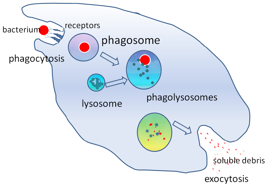
For instance number of phagocytic cells such as macrophages and neutrophils engulf large particles following the process of phagocytosis during which the single membrane phagosome is formed which can then fuse directly with the lysosome which forms the phagolysosome.
In case of internalization of the fluid substances by the process of pinnocytosis, the endocytosed substances eventually end up in lysosomes where they are degraded.
Autophagy, on the other hand begins with the compartmentalization with the formation of phagophore which then expands into a double membrane bound autophagosome surrounding a portion of the cytoplasm. This autophagosome can then fuse with an endosome.
The product of this fusion is called as amphisome. The amphisome then fuses with the lysosomes where the acid hydrolases are present for the process of degradation. There are some lysosomes which participate in the process of exocytosis.
Due to this process the undigested contents are eliminated from the cell. These pathways acta s minor pathway for various cells, but there are certain cells in which the lysosomes have acquired the specialised machinery in order to fuse with the plasma membrane which aids in the process of exocytosis.
Cells such as melanocytes produce and store melanin pigments in their lysosomes called as melanosomes. These pigments containing melanosomes release their pigment into the extracellualr space of the epidermis by exocytosis.
Final Words
I hope that now you have a clear idea about Lysosomes, its structure and functions. If you have any suggestion or feedback, please leave a comment.
You can also read Mitochondria- The Powerhouse of the Cell.
I will surely foreward this post to all of my pals! Its very first-class and a very respectable check out!